Microscopy
Vision Solutions for conventional light and fluorescence microscopy
Vision technology in microscopy enables the precise examination of different structures and functions. Our cameras deliver the highest image quality in real time, with excellent color reproduction and a high dynamic range. The result: reproducible, detailed observation and analysis of the finest structures.
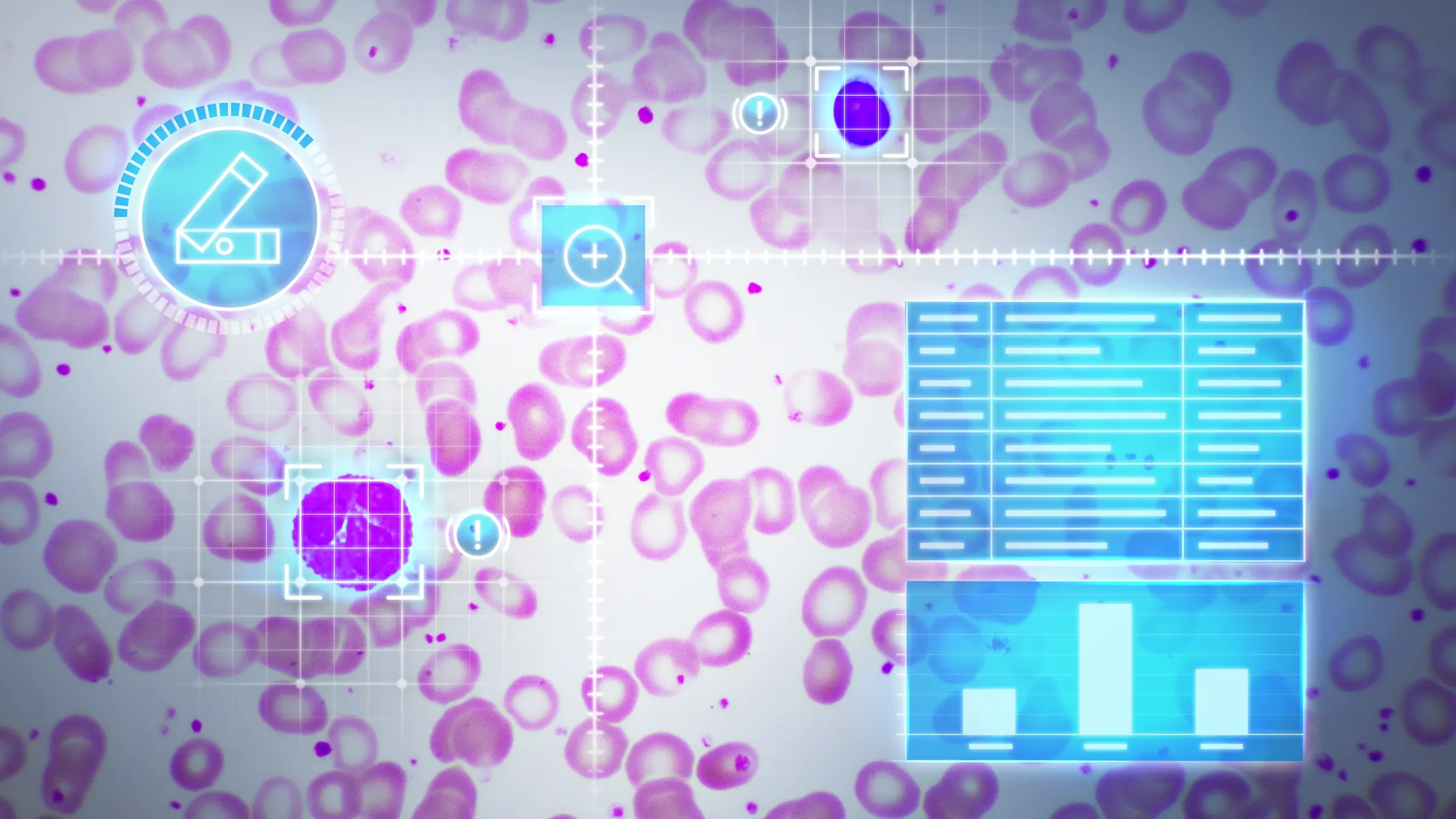
Best image quality and color reproduction
Basler cameras produce excellent images - whether they operate completely automatically or are individually adjustedCustomizable hardware and software
Our project teams work with you to design tailored solutions that meet your lab’s specific needs.Industry-leading reliability
Basler cameras have one of the lowest failure rates in the industry — built for continuous, 24/7 operation.Long-term product availability
Future-proof your system design with products that stay available and supported over time.
Typical applications in the pharmaceutical industry
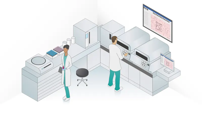
Vision solutions for image acquisition and processing are an integral part of microscopy. Light microscopy, including fluorescence microscopy, enable numerous applications in scientific, biomedical, and clinical laboratories. These include: histology, cell biology, hematology, and medical microbiology.
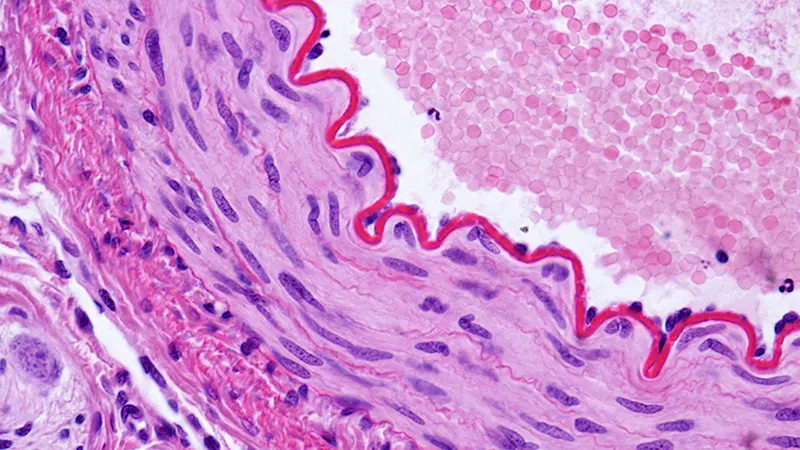
Conventional light microscopy
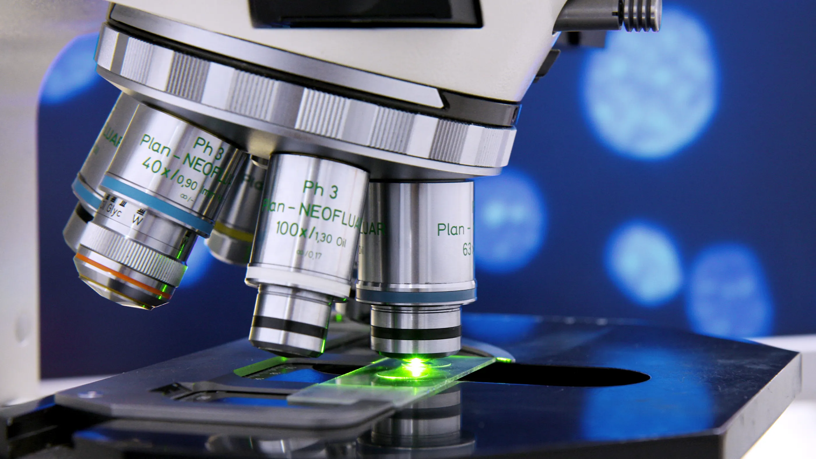
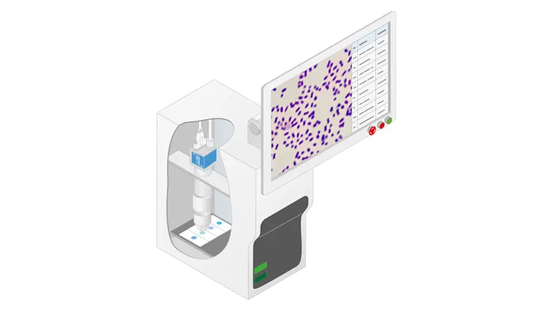
Many routine microscopic applications are based on light microscopy, using different illumination and contrasting techniques. Cameras are a central component of these microscopy applications, they help track, record, analyze, and archive live images. The cameras used must reliably deliver high resolution and sharp images with accurate color fidelity.
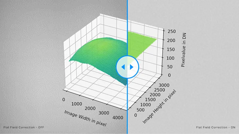
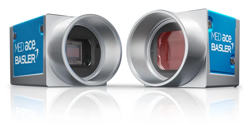
Light microscopy applications benefit from MED Feature Set "Brilliant Image"
"Brilliant Image" is one of the MED Feature Sets that distinguish our Basler MED ace cameras. Learn about two features that help deliver the maximum image quality for your application.
Fluorescence microscopy
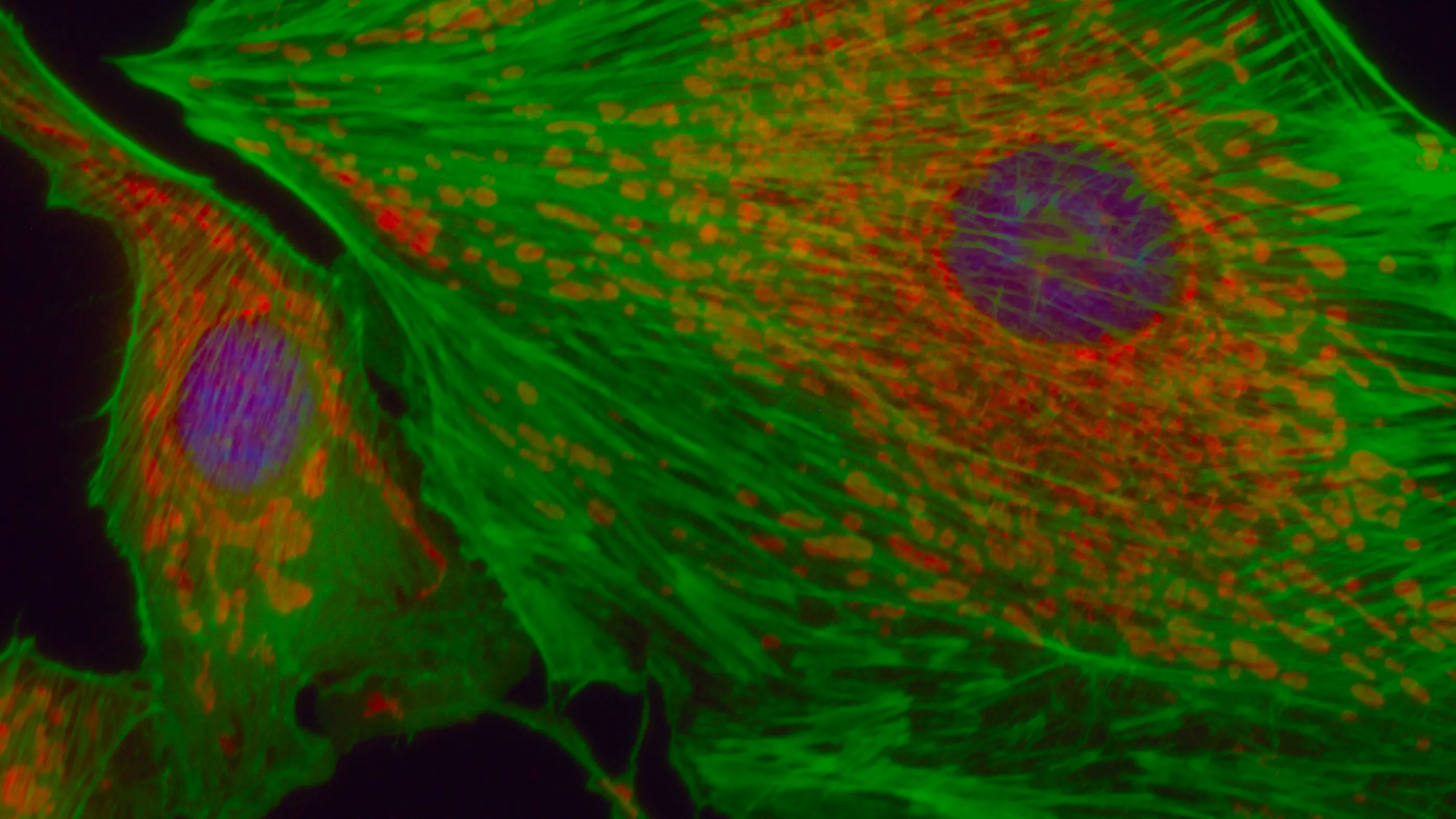
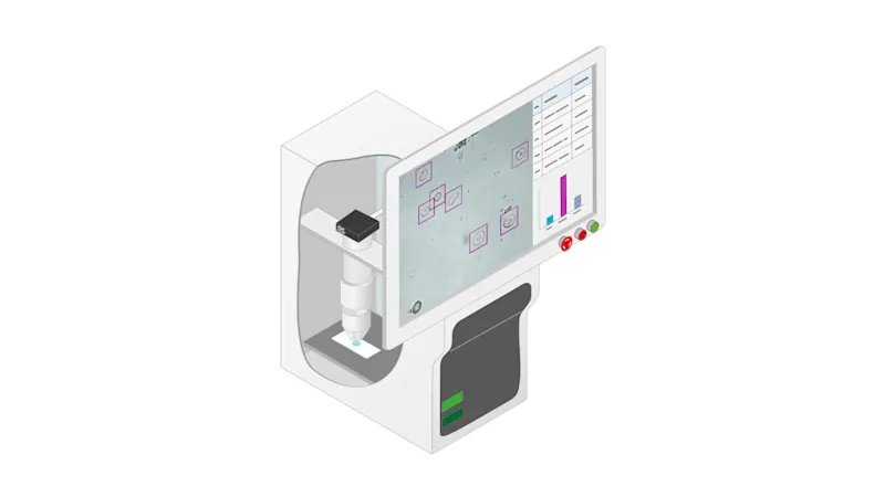
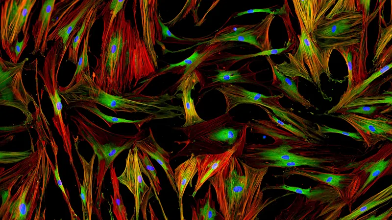
A certain wavelength of light excites fluorescent dyes, causing them to glow and allowing the light emission to be detected. The smallest cellular and even molecular structures can be distinguished with fluorescence imaging. The camera technology is critical for application success: with minimal noise, a high dynamic range, and high light sensitivity, Basler cameras are ideally suited for fluorescence microscopy applications.
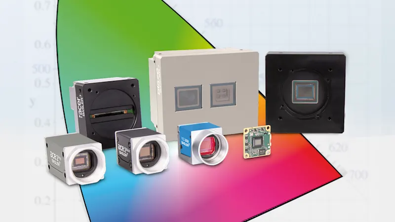
Color calibration in Medical & Life Sciences
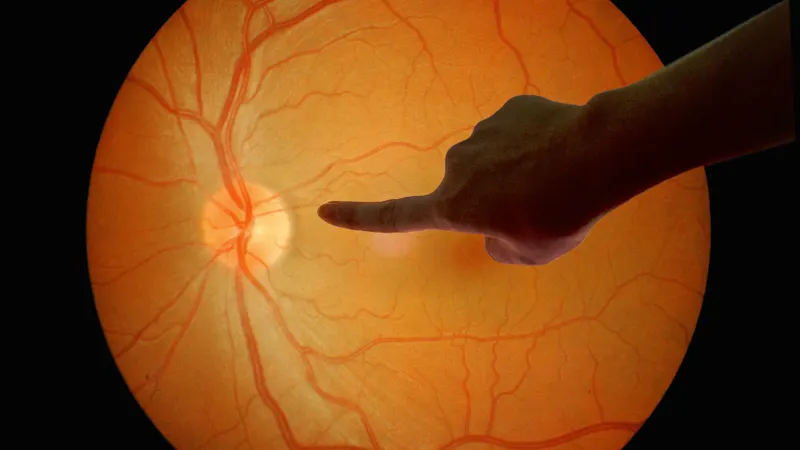
Digitally captured images contribute significantly to the diagnostic and therapeutic process, especially in the fields of microscopy, ophthalmology, dermatology, and surgery (including both medical imaging systems and procedures). In this context, color is often a decisive criterion for assessing whether a structure is healthy or pathological. Color fastness is therefore of utmost importance.
Application examples in microscopy
There are several options that can be used to save time and money for a wide variety of applications. Our customized solutions can help.
Most popular products
For efficient and reliable machine vision applications in this industry, the following Basler products are often the best choice:
